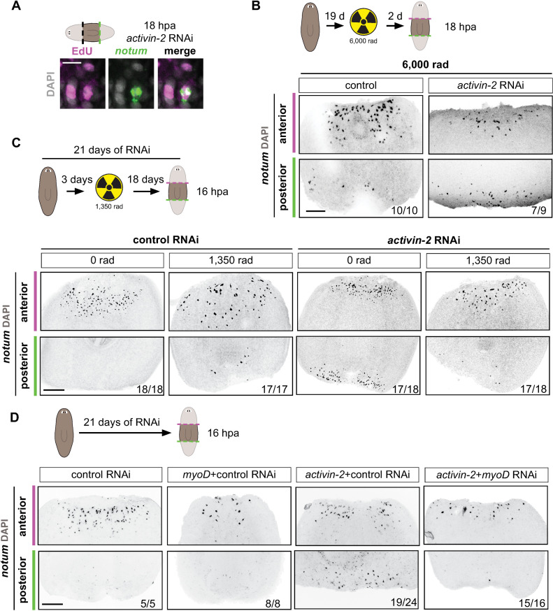
Fig 5. activin-2 promotes the polarized response of newly specified longitudinal muscle fibers to wound orientation.
(A) EdU incorporation in notum+ cells at posterior-facing wounds in an activin-2 RNAi animal. Lower magnification in S5A. (B) Top: cartoon shows the experimental design. Bottom: FISH shows notum expression at 18 hours post-amputation 21 days after the first RNAi feeding in lethally irradiated (6,000 rads) animals. (C) Top: cartoon shows the experimental design. Bottom: FISH shows notum expression at 18 hours post-amputation 21 days after the first RNAi feeding where a sublethal dose of irradiation (1,350 rads) was given 19 days prior to amputation. (D) Top: Cartoon showing experimental design. Animals were fed every 3 days. Animals were fed either control or activin-2 dsRNA for feeding 1, 2, 3, 5, and either myoD or control for feeding 4. Bottom: FISH shows notum expression at 18 hours post-amputation 21 days after the first RNAi feeding. Magenta, anterior-facing wounds; green, posterior-facing wounds. All images are anterior up. Scale bars, 200 μm, except for high magnification panels in where they represent 10 μm.

