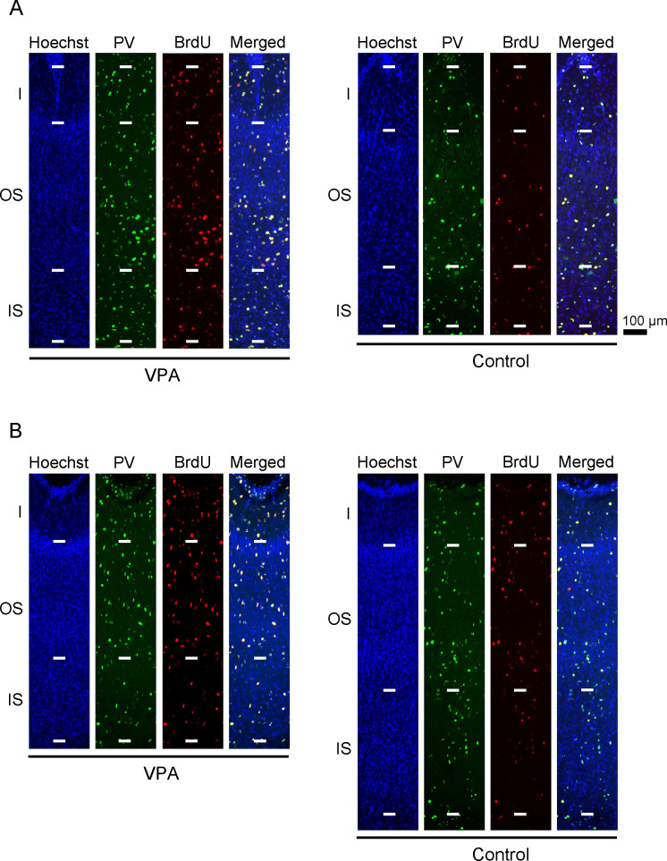
Fig 3. Parvalbumin neuron immunofluorescence with BrdU labeling and Hoechst staining in sulcal floors of the cerebral cortex of VPA-treated and control ferrets at PD 20.
(A) Cortical depth of the rostral sylvian sulcus (rsss) floor. (B) Cortical depth of the lateral sulcus (ls) floor. The rsss and ls were selected as representative sulci with cortical floors that were thickened or thinned by neonatal VPA exposure, respectively. PV, parvalbumin.

