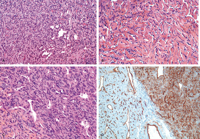
Figure 1.

A,B. Microscopic features of a solitary fibrous tumor: characteristic biphasic pattern: cellular areas with staghorn vessels (A, HES X 25) and pseudokeloidal collagenous areas (B, HES X 25). C,D. Microscopic features of a hemangiopericytoma: highly cellular tumor made of oval cells arranged around vessels (C, HES X 40). Heterogenous expression of CD34 by immunohistochemistry (D, X 25).
