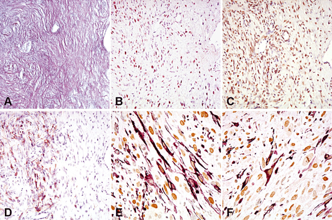
Figure 2.

Consecutive sections of the mesenchymal tumor area of a gliosarcoma co‐expressing reticulin (A), Slug (B) and Twist (C). Note GFAP expression (D) in a focal area. Double stainings show co‐expression of GFAP (purple, cytoplasms) and Slug (brown nuclei) (E), or GFAP (purple, cytoplasms) and Twist (brown nuclei) (F) at the single‐cell level.
