
Figure 1.
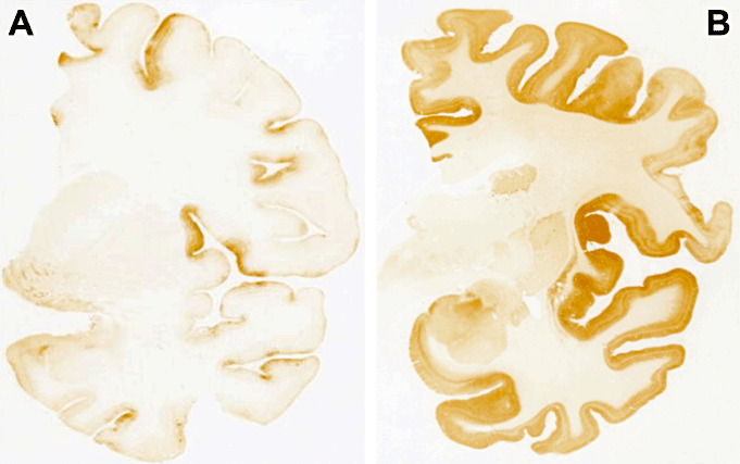
Prion protein (PrP) immunohistochemistry (3F4 antibody) of patient 1 (A) and patient 2 (B). Patient 1 showed faint PrP deposition mainly involving the cerebral cortex with focal areas of increased intensity such as the middle frontal gyrus and the superior part of the insula. Patient 2 displayed a diffuse, synaptic‐like pattern of PrP deposition that was intense in the cerebral cortex and weaker in striatum, closely resembling a typical MM1 case.