
Figure 2.
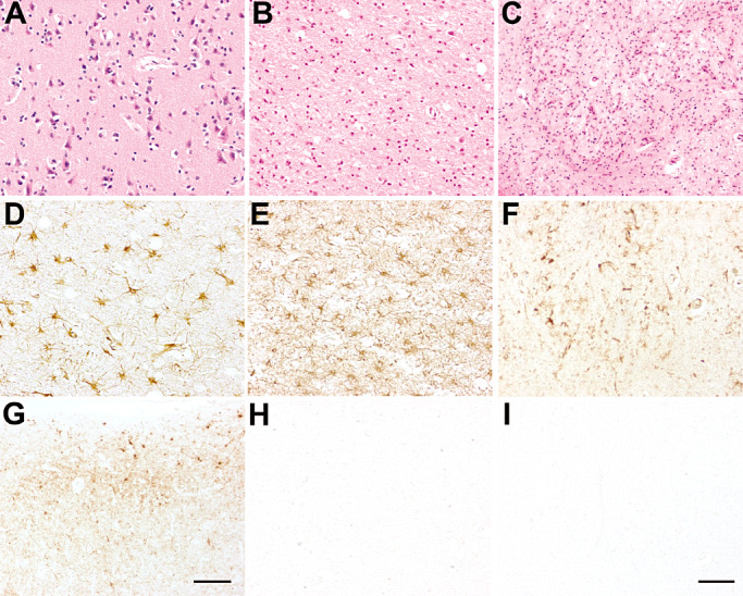
Patient 1: neuropathological findings in the cerebral cortex (A,D,G), dorsal nuclei of the thalamus (B,E,H) and olivary nucleus (C,F,I). Hematoxylin‐eosin (H&E) staining showed mild spongiform changes in the cerebral cortex (A), and severe neuronal loss in the thalamus (B) and olivary nucleus (C). Astrogliosis [glial fibrillary acidic protein (GFAP) immunostaining] was moderate in the cerebral cortex (D) and severe in the thalamus (E) and olivary nucleus (not shown), and was accompanied by marked microglial activation (F, olivary nucleus, CR3/43 immunostaining). Prion protein (PrP) immunoreactivity (3F4 immunostaining) exhibited a synaptic pattern with sub‐pial focal deposits in the cerebral cortex (G) but spared the thalamus (H), cerebellum (not shown) and brainstem (I, olivary nucleus). Scale bar in I = 5 µm. A, B, C, D, E, F, H, I are at the same magnification. Scale bar in G = 15 µm.