
Figure 3.
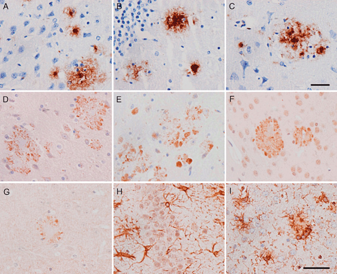
Main neuropathological findings in APP/PS1 mice aged 6 months. β‐amyloid plaques in the neocortex (A), hippocampus (B) and entorhinal cortex (C). Amyloid plaques are surrounded by dystrophic neurites immunoreactive with mitochondrial porin/voltage‐dependent anion channel (VDAC), as revealed with Abcam (D) and Calbiochem (E), antibodies and ubiquitin (F). BACE 1 is also expressed in association with β‐amyloid plaques (G). Hypertrophic astrocytes, as revealed with GFAP antibodies (H) and microglia stained with Lycopericum esculentum lectin (I), are also observed in the vicinity of plaques. Paraffin sections slightly counterstained with hematoxylin. A–G, bar in C = 25 µm; H, I, bar in I = 40 µm.