
Figure 5.
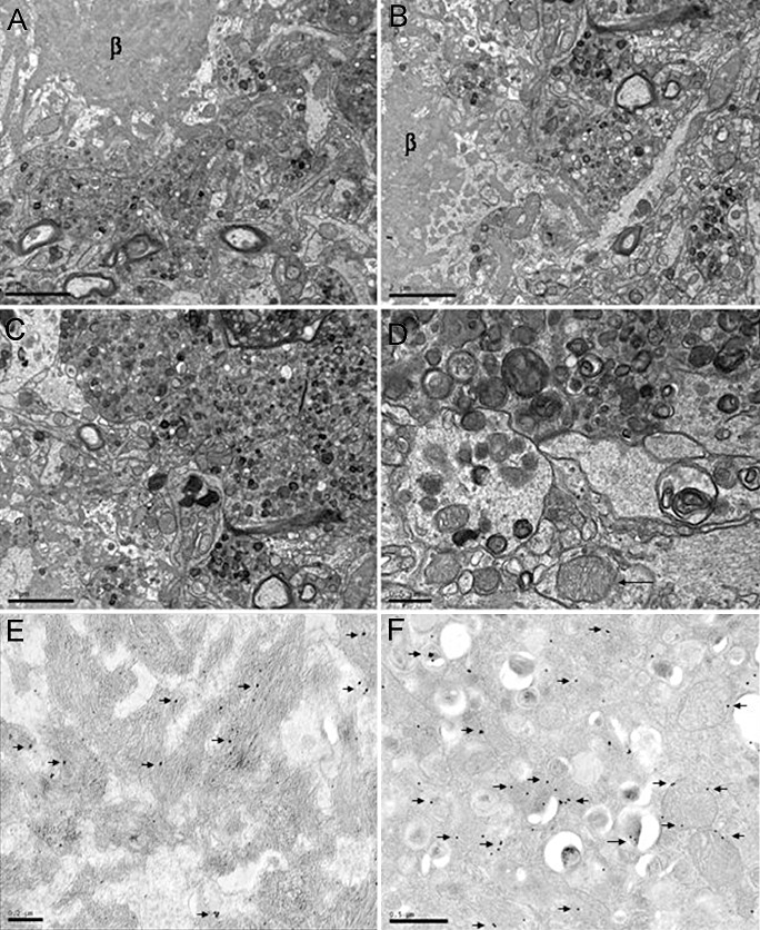
A–C. Electron microscopy of β‐amyloid plaques showing a central, radiating core of amyloid (β) surrounded by aberrant huge neurites filled with altered mitochondria, polymorphous inclusions and vesicles. APP/PS1 mice aged 12 months. A–C, bar = 2 µm; D, bar = 0.5 µm. D,F. Immunoelectronmicroscopy showing immunogold particles (arrows) decorating β‐amyloid in the core of a plaque (D) and VDAC in the membrane of mitochondria and polymorphous inclusions in aberrant neurites surrounding β‐amyloid deposits (F). APP/PS1 mice aged 6 months. E, bar = 0.2 µm; F, bar = 0.5 µm.