
Figure 7.
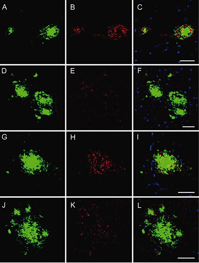
Double‐labeling immunofluorescence and confocal microscopy showing altered phospho‐tau deposition (red, B, E, H, K) in association with increased neuroketal immunoreactivity in plaques (green, A, D, G, J). C, F, I, L: merge. Nuclei are stained in blue (DRAQ5). APP/PS1 mice aged 6 and 9 months. A–F, bar = 25 µm; G–L, bar = 25 µm.