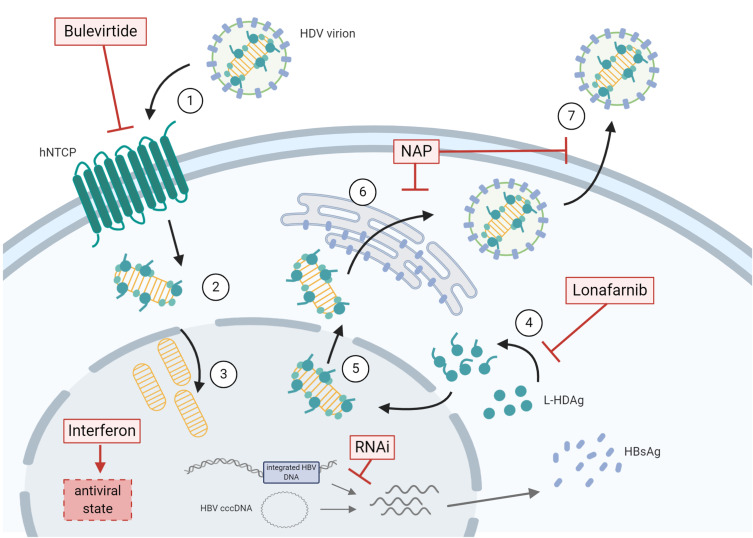
Figure 1.
Therapeutic approaches for the treatment of chronic HDV infection are depicted in relation to the viral life cycle. (1) HDV virions attachment to heparan sulfate proteoglycans and binding of L-HBsAg pre-S1 region to the HBV/HDV specific receptor, hNTCP. Viral particles enter the cell through endocytosis and the viral ribonucleoprotein (RNP) is released in the cytoplasm. (2) Viral RNP translocation to the nucleus. (3) HDAg mRNA transcription and replication of HDV RNA. (4) L-HDAg contains a prenylation site that is farnesylated by a cellular farnesyltransferase before being re-translocated to the nucleus. (5) Both forms of HDAg interact with the newly synthesized genomic RNA to form new viral (RNP) that are exported to the cytoplasm. (6) Viral RNPs interact with the cytosolic part of HBsAg at the endoplasmic reticulum surface inducing their envelopment. (7) HDV virions are secreted form the infected cell. The different steps targeted by antiviral treatments are depicted. HBV co-infection is represented by presence of HBV cccDNA and integrated HBV DNA. The figure was created with BioRender.com.
Abbreviations: cccDNA, covalently closed circular DNA; HBsAg, hepatitis B surface antigen; HDV, hepatitis D virus; hNTCP, human sodium taurochlorate co-transporting polypeptide; L-HDAg, large hepatitis D antigen; NAP, nucleic acid polymers; RNAi, RNA interference compounds.

