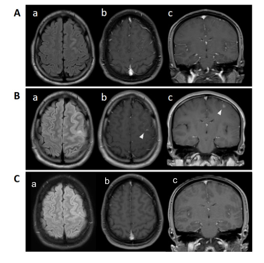
Figure 1. MRI of the brain with and without contrast. MRI of the brain after initial seizure (A), axial FLAIR showing area of increased signal within the left frontal gyrus (A.a), with no enhancement on post-contrast axial (A.b) and coronal (A.c). During the second admission (B), axial FLAIR showing increased signal within cortex and subcortical white matter (B.a), and area of enhancement (arrowhead) within the same region on post contrast axial (B.b), and coronal (B.c). On follow-up imaging (C), disappearance of hyperintense signal (C.a).

