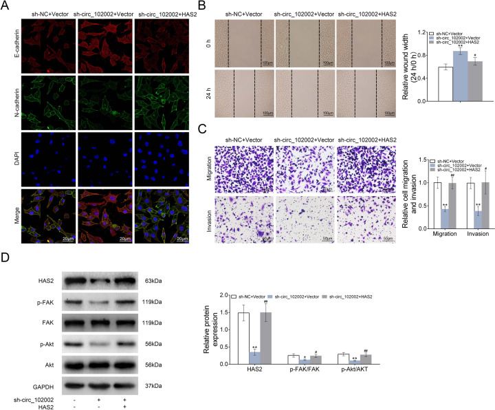
Fig. 6. Hsa_circ_102002/HAS2 promoted the epithelial–mesenchymal transformation and migration of PTC cells through regulating FAK/Akt passway.
a Representative images of E-cadherin and N-cadherin in sh-NC + Vector, sh-circ_102002+Vector, and sh-circ_102002 + HAS2 TPC-1 cells, as determined using IF. (red, E-cadherin; green, N-cadherin; blue, DAPI). b Wound scratch healing assay of sh-NC + Vector, sh-circ_102002+Vector, and sh-circ_102002 + HAS2 TPC-1 cells. Quantification of the wound-healing assay was shown as histograms. c Representative migration and invasion assay images of sh-NC + Vector, sh-circ_102002+Vector, and sh-circ_102002 + HAS2 TPC-1 cells. The migrated cells were quantified and shown as histograms. d Protein expression levels of HAS2, p-FAK, FAK, p-Akt, and Akt in sh-NC + Vector, sh-circ_102002 + Vector, and sh-circ_102002 + HAS2 TPC-1 cells, as determined using western blotting. Bands were quantified and shown in histogram (mean ± SEM, *p < 0.05, **/##p < 0.01).

