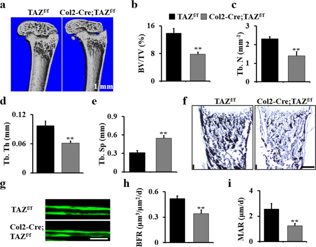
Fig. 3. Deletion of TAZ in chondrocytes impairs bone development.
a Representative micro-CT reconstructions of the femurs of Col2-Cre;TAZf/f mice and controls at 1 month. Scale bars, 1 mm. b–e Histomorphometric analysis of bone parameters in the femur of Col2-Cre;TAZf/f and control mice (n = 5 mice per group). Bone volume fraction (BV/TV) (b); trabecular thickness (Tb.Th) (c); trabecular number (Tb.N) (d); trabecular spacing (Tb.Sp) (e). f Representative von Kossa staining of terminally differentiated hypertrophic chondrocytes in the tibiae of mice at E18.5 (n = 3 mice per group). Scale bars, 75 μm. g–i Calcein double labeling in tibia of 2-month-old Col2-Cre;TAZf/f mice and controls. The mice were injected with calcein twice with an interval of 7 days. The mice were sacrificed 1 day after the second injection. The tibia bones were embedded, sectioned, and the images were taken using a microscope. h Bone formation rate (BFR); i mineral apposition rate (MAR). n = 5 mice per group. The experiment was repeated three times independently. Error bars were the means ± SEM of triplicates from a representative experiment. **P < 0.01.

