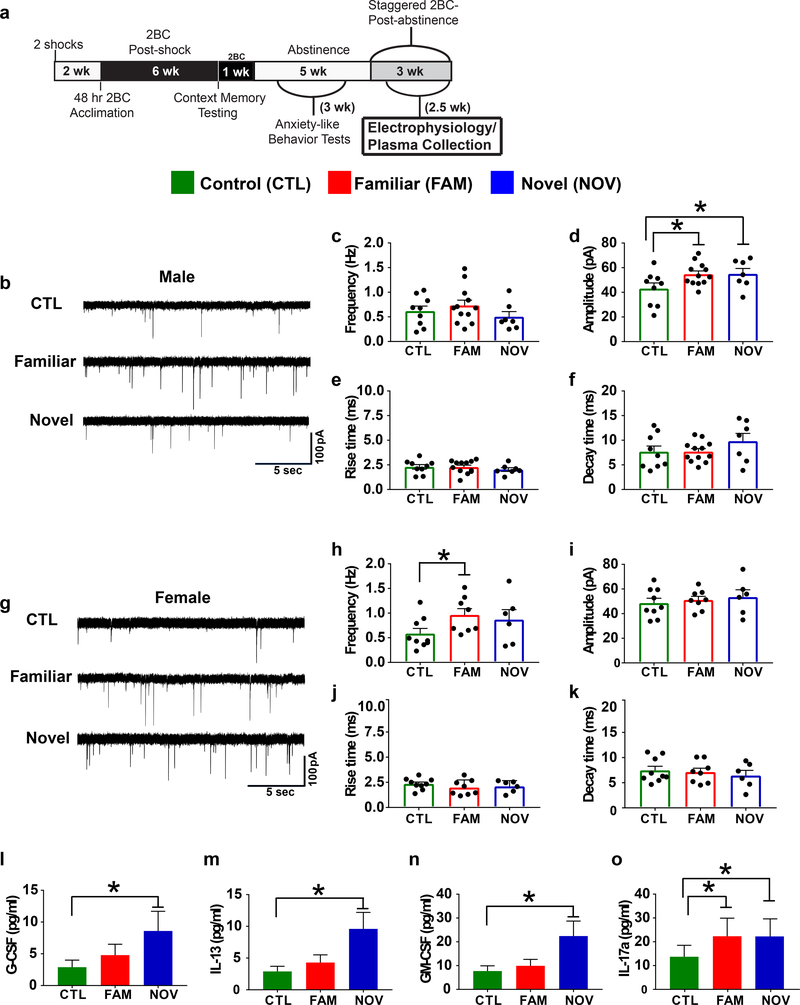
Fig. 4. Stress increased central amygdala (CeA) inhibitory GABAergic transmission and elevated peripheral cytokines.
(a) Timeline showing when physiological endpoints were measured (electrophysiology and plasma collection for cytokine analysis). (b-k) We examined CeA miniature inhibitory postsynaptic currents (mIPSCs). Representative mISPCs for males (b) and females (g) performed 24-hr into abstinence, 104–123 days post-first stressor (n=3–4 rats/group, # cells/group=6–12 (see scatter plots). (d) Within-sex planned comparisons demonstrated that among males, FAM (P=0.025) and NOV (P=0.044) stress significantly increased mISPC amplitude. (h) Conversely, FAM females exhibited increased mIPSC frequency (P=0.034). Frequency was not affected in males (c) and amplitude was not affected in females (i), andthere were no differences in rise (e,j) or decay (f,k) times in either sex (all P’s>0.05, planned comparisons). (i-o) Graphs showing backlog transformed cytokine and chemokine concentrations with Luminex plate (n=2) as a covariate (n=7–8 rats/group; 1 FAM and 1 CTL male removed as outliers, n=7 NOV males). Least significant difference analyses indicated that NOV males showed significantly elevated plasma concentrations of G-CSF, IL-13 and GM-CSF compared with controls (P’s≤0.04), while plasma IL-17a levels were elevated in both NOV (P=0.034) and FAM (P=0.041) males. *P<0.05, within-sex planned comparisons. All data are shown as mean±SEM.

