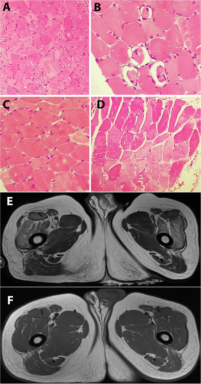
Fig. 2.
Muscle histopathology and MRI of upper thigh axial sections. (a-d) Muscle histopathology sections sampled from quadriceps femoris muscle stained with hematoxylin and eosin, showed variation in muscle fiber size with predominantly spherical shape myosites and multifocal and discrete myosite degeneration lacking infiltration of inflammatory cells. (e) T2 weighted MRI section of the upper thigh done on day 20, showed marked hyperintensity in both the quadriceps muscles. (f) Repeat T2-weighted MRI of the same section of the thigh muscles done after 48 days of the 1st MRI shows both the quadriceps muscles appear normal

