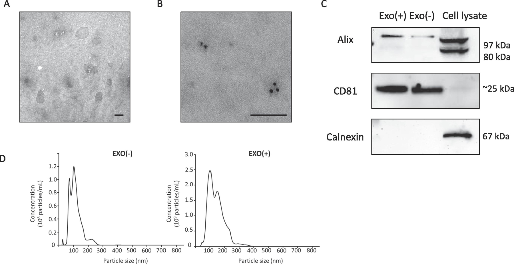
Figure 1. Characterization of sEVs derived from RAW 264.7 cells.

(A) Transmission electron microscopy (TEM) images of sEVs purified from culture media of RAW 264.7 cells showing size range and integrity (scale bar = 100 nm). (B) Immunogold labeling for CD81 (scale bar = 100 nm). (C) Western blot analysis of sEVs lysate from unstimulated (Exo−) or stimulated (Exo+) and RAW 264.7 cell lysate indicates that two commonly detected sEVs proteins, Alix and CD81 are present in sEVs. The negative marker Calnexin is absent in sEVs but present in whole cell lysate. The absence of negative marker suggests purity of our sEVs preparations. (D) Nanoparticle tracking analysis (NTA) of sEVs showed Exo(−) particles with a mean diameter of 113.6 ± 7.9 nm and a particle concentration of 8.6 x 10⁸ ± 9.8 x 10⁷ particles/ml and for Exo(+) the mean diameter was 143.3 ± 0.8 nm and a particle concentration 2.6 x 10⁹ ± 3.2 x 10⁷ particles/ml. On average we found 1 µg sEVs protein equals to 1 x 10⁹ particles.
