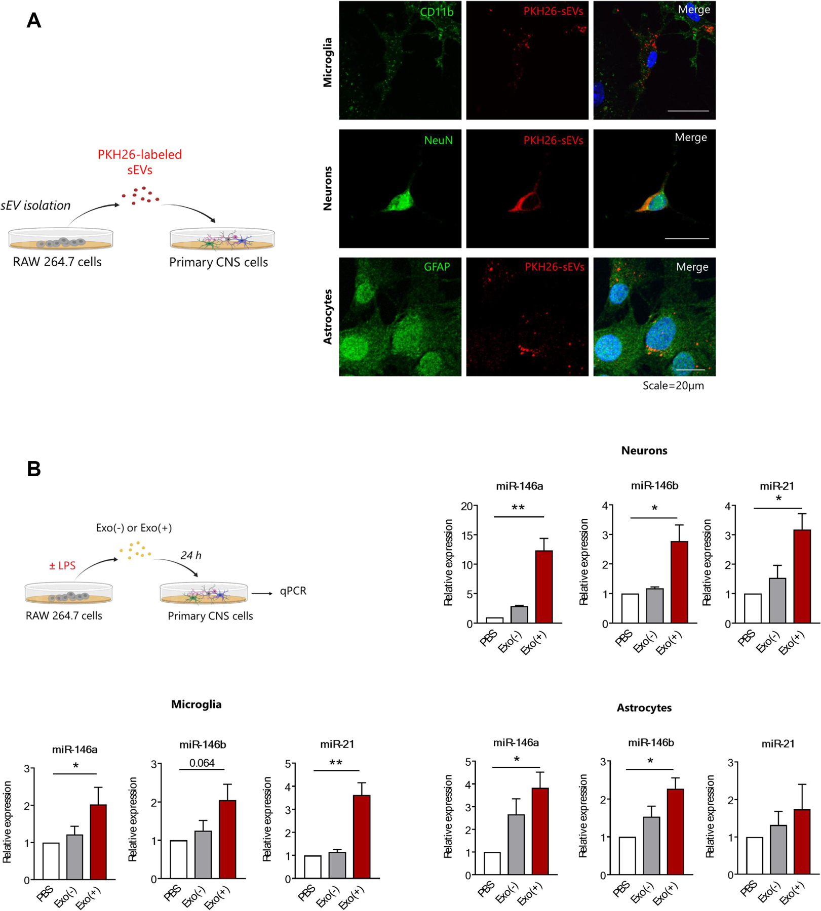
Figure 3. Uptake of sEVs transferred miRNAs to primary microglia, neurons, and astrocytes.

(A) Experimental design and representative images of immunostaining showing sEVs are internalized by primary microglia, neurons, and astrocytes. PKH26 labeled sEVs (red) are taken up by cells labeled for specific markers; primary microglia (anti-CD11b), neurons (anti-NeuN), and astrocytes (anti-GFAP). Scale bar = 20 µm. (B) Schematic of in vitro experiment and qPCR analysis of miRNAs in recipient cells incubated with sEVs. The sEVs isolated from RAW 264.7 cell conditioned media from naïve (Exo(−) or after 24 h stimulation with 1µg LPS (Exo(+) were incubated with primary cells for 24 hours. Exo(+) sEVs treatment increased relative levels of miR-146a, miR-146b and miR-21 in recipient cells. Expression of miR-146a, miR-146b and miR-21 were normalized to U6 snRNA (n=3). All data shown as mean ± SEM and analyzed with one-way ANOVA *p < 0.05, **p <0.01.
