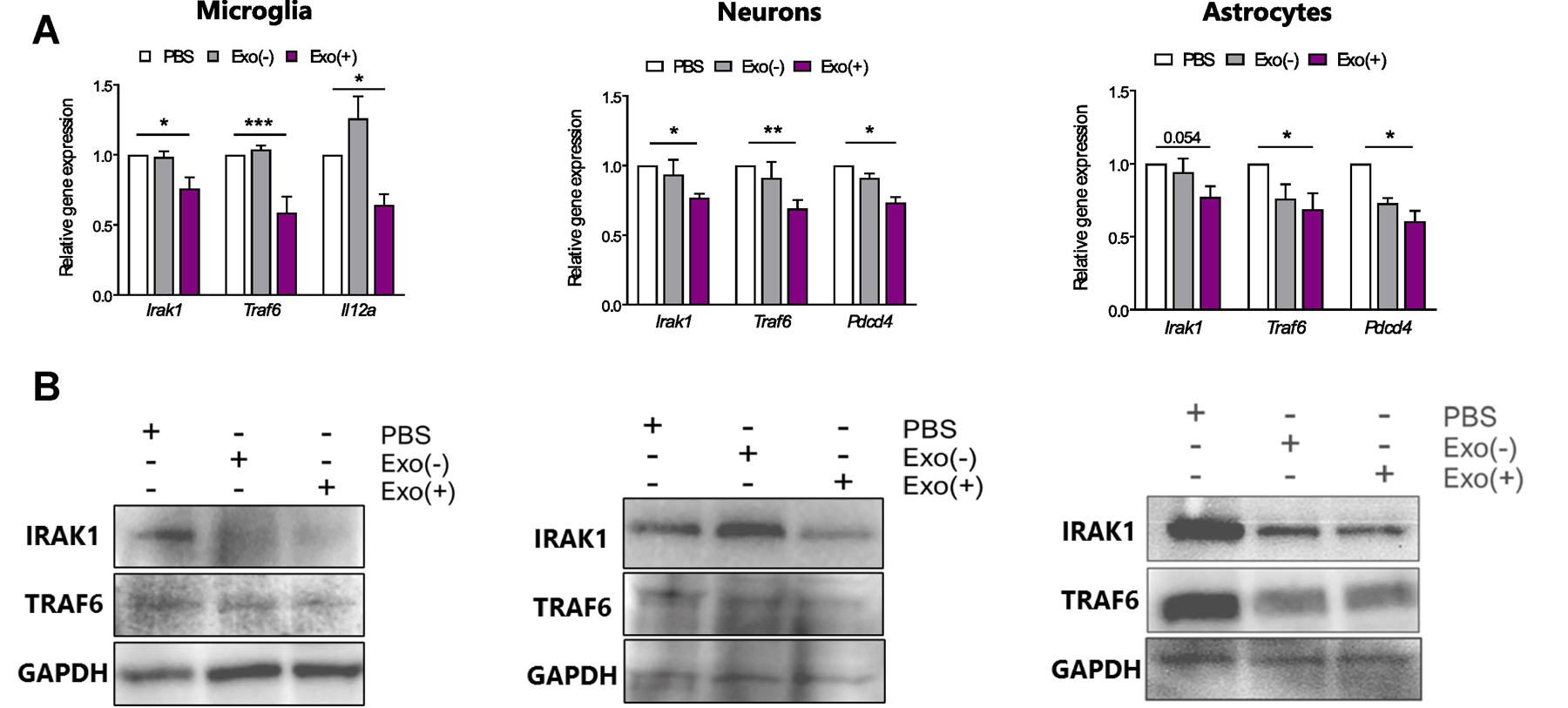
Figure 4. sEV-derived miRNAs are functional and decrease their target mRNA and protein in in primary microglia, neurons and astrocytes.

Exo(+) treatment reduced relative mRNA and protein levels of genes targeted by Exo(+) enriched miRNAs in all cell types. (A) qRT-PCR was used to measure relative mRNA levels of miR-146a, miR-146b, and miR-21 targets, Irak-1, Traf6, and Il12a or Pdcd4 respectively, in microglia, neurons and astrocytes following 24 h incubation with sEVs (n=3). Data shown are mean ± SEM. Statistical analysis was determined by one-way ANOVA *p < 0.05, **p < 0.01, ***p < 0.001. (B) Representative western blots of IRAK-1, TRAF6 and GAPDH in sEVs treated microglia, neurons and astrocytes.
