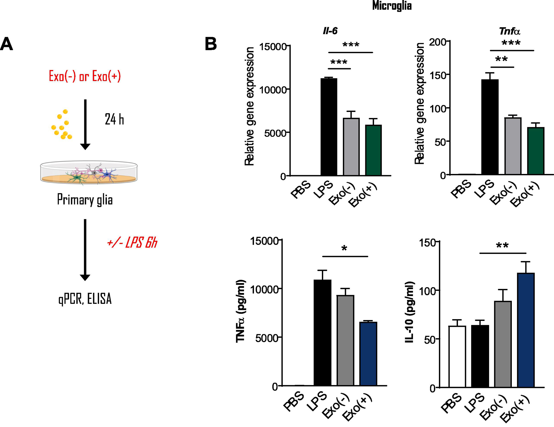
Figure 5. Pre-incubating microglia with RAW 264.7-derived sEVs reduced LPS-induced proinflammatory gene expression.

Exo(−) and Exo(+) treatment decreased mRNA levels of Tnf and Il6 in primary microglia stimulated with LPS as determined by qPCR. ELISA for secreted cytokines showed that Exo(+) treatment decreased TNF and increased anti-inflammatory IL-10 secretion from these cells, (n=3). All data shown as mean ± SEM and analyzed with one-way or two-way ANOVA followed by Dunnett’s post-test *p < 0.05, **p <0.01, ***p <0.001.
