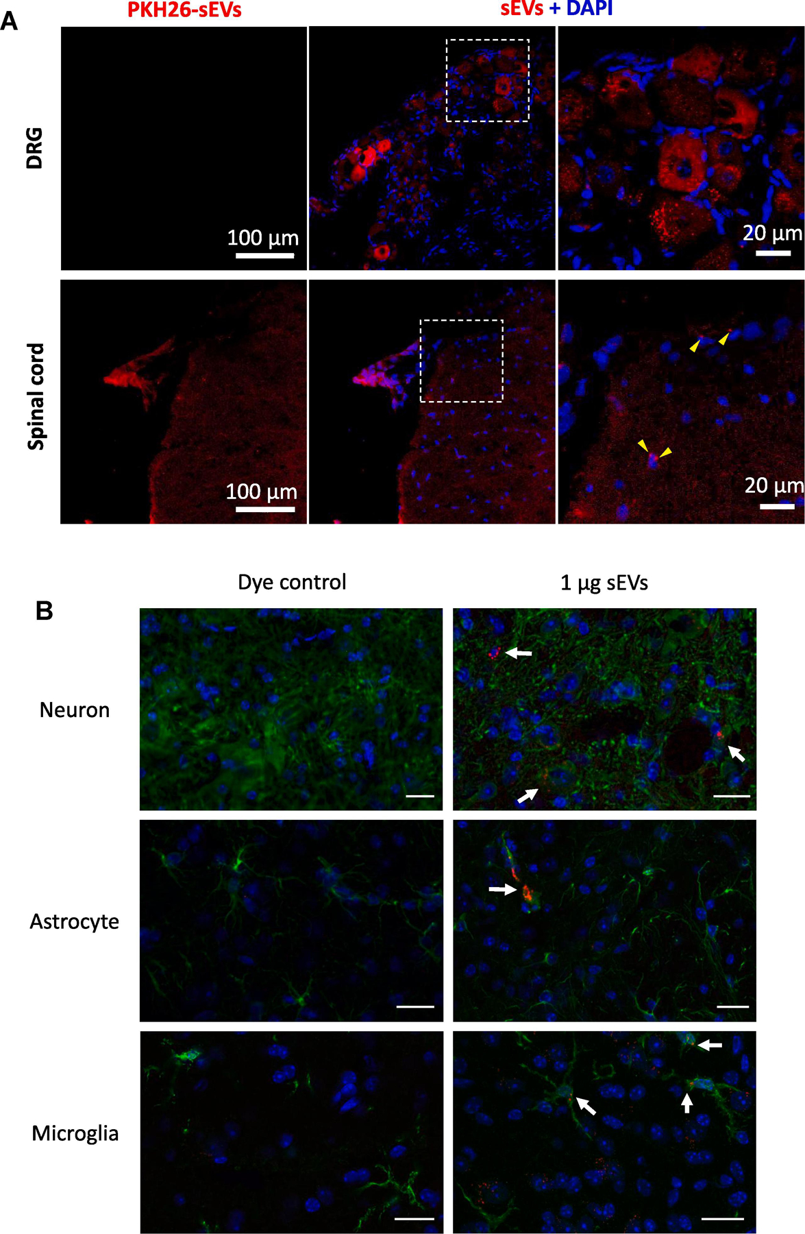
Figure 6. Uptake of intrathecally injected sEVs in mice.

(A) Intrathecal (i.t.) injection of sEVs isolated from conditioned media of RAW 264.7 cells labeled with PKH26 showed uptake by dorsal root ganglion (DRG) (top) and spinal cord (SC) (bottom) (1 μg in 10 μl PBS). Red PKH26, Blue DAPI, Scale bar =100µm. (B) Uptake of sEVs in vivo by neurons, astrocytes and microglia in vivo. One µg PKH26 labeled sEVs were injected intrathecally and after 18 h of sEVs injection, mice were perfused with 4% PFA. Spinal cord was isolated and 30 µm section were stained for MAP2A as neuronal marker, GFAP as astrocytic marker, and Iba1 as microglial marker. Nuclei were stained with DAPI and PKH26 labeled sEVs are shown in red. A dye control was included for all cell types. sEV uptake was observed under a confocal laser scanning microscope. Scale bar = 20 µm. Arrows show the labeled sEVs uptake.
