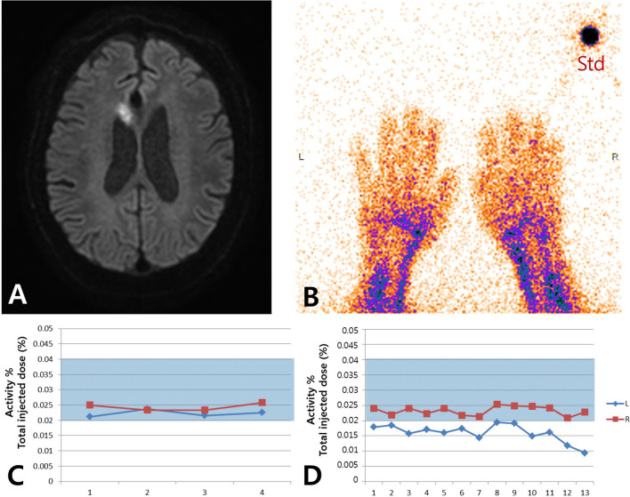
Figure 2.
Modified Raynaud scan of impaired vasoreactivity case. (A) High signal intensity in right anterior cerebral artery territory on a diffusion-weighted image. (B) Scan for both hands and standard source. (C) Same blood amount in both hands. Activity % of the total injected dose means the percentage of total blood. (D) Impaired vasoreactivity in the left hand under thermal stress. The amount of blood in the right hand was stable during the scan. The shaded area in Fig. 1 (C,D) represents the reference range obtained from 10 normal volunteers. R: right; L: left; Std: standard.

