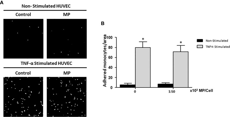
Figure 5.
Effects of Membrane Particles on monocyte adhesion to TNFα-activated HUVEC. HUVEC were stimulated with TNFα and treated with MP at ratio 1:50,000. Subsequently, CFSE-labeled monocytes were added during 1h. (A) Representative fluorescent microscopy pictures show the adhered monocytes (white dots) to the HUVEC monolayer in non-stimulated and TNFα stimulated conditions. (B) Quantitative results of the monocyte adhesion assay analyzed by imageJ. No significance difference respect to the respective control (Non-Stimulated, and TNFα Stimulated HUVEC) was observed when MP were added *p < 0.05 compared to Non-Stimulated HUVEC.

