Figure 6.
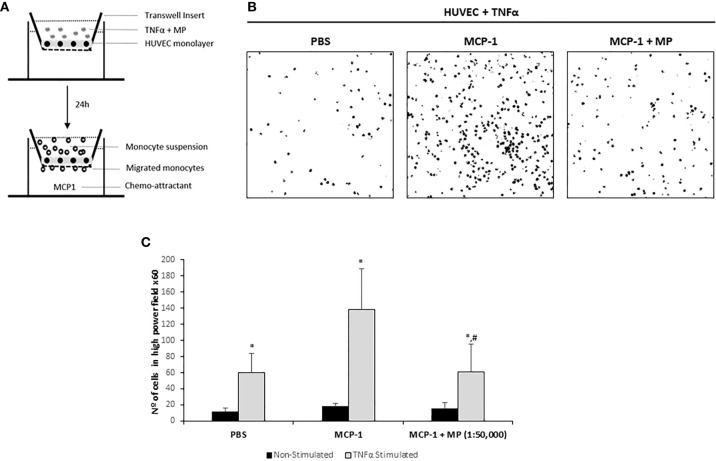
Effect of Membrane Particles on migration of monocytes through a monolayer of TNFα-activated HUVEC. (A) Schematic representation of the transmigration assay. HUVEC were seeded on transwell inserts until confluency. The monolayer of cells was treated with TNFα and 1:50,000 MP for 24h. Then, 1x10⁵ isolated monocytes were added during 2h with addition of the chemo-attractant MCP-1 in the bottom well. Three pictures from randomly selected areas of the transwell were taken for the quantification. (B) Representative confocal microscopy images of the negative control (no MCP-1), positive control (MCP-1) and the MP treated group analyzed by ImageJ. (C) Quantitative results of the transmigration assay. Data represent means ± SD of the number of transmigrated monocytes. *p < 0.05 respect to Non-Stimulated HUVEC. #p < 0.05 respect to TNFα stimulated HUVEC non treated with MP in the MCP-1 group.

