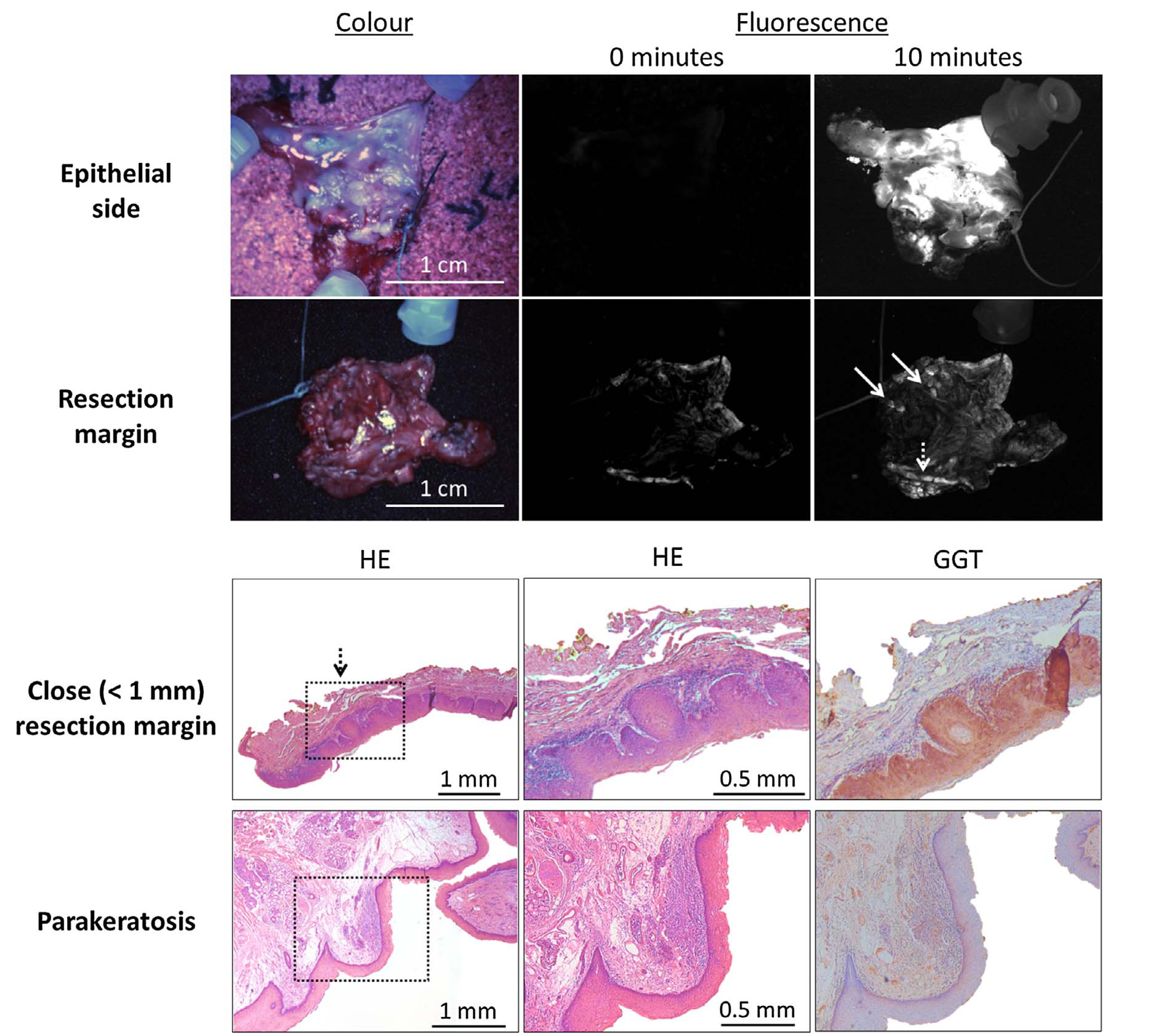
Fig. 5.

Example of resection margin assessment with gGlu-HMRG and the clinical Artemis imaging system (upper two panels). Ten minutes after spraying on the tissue, several spots became fluorescent in the resection margin (arrows). In one patient with squamous cell carcinoma, focal expansion of a carcinoma in situ < 1 mm from the resection margin was diagnosed with fluorescence imaging (dashed arrow). The epithelial side was used as a positive control.
