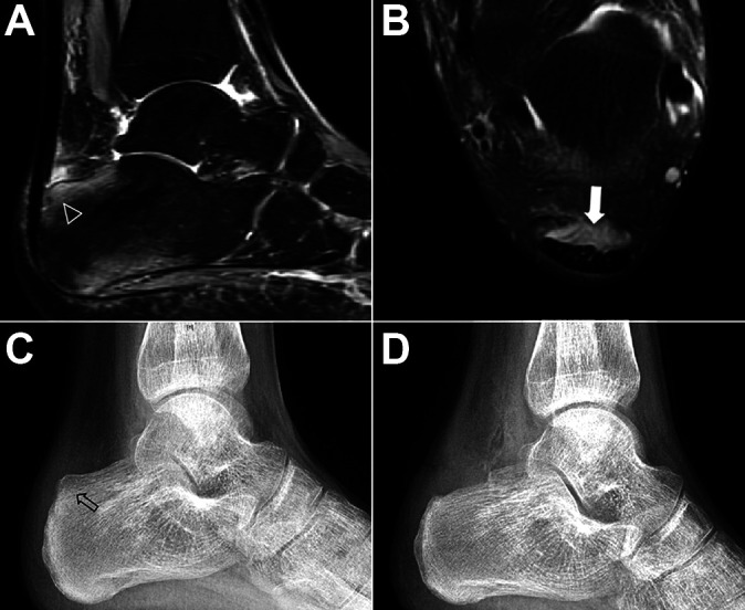
Figure 3.

Endoscopic procedure for Haglund syndrome. (A and B) Retrocalcaneal impingement as visualized on magnetic resonance imaging. (A) Sagittal view with bone marrow edema (arrowhead). (B) Axial view with retrocalcaneal bursitis (white arrow). (C and D) Lateral ankle radiographs (C) with enlargement of posterosuperior calcaneal tuberosity with Haglund deformity (arrow) and (D) after posterosuperior calcaneal tuberosity was excised.
