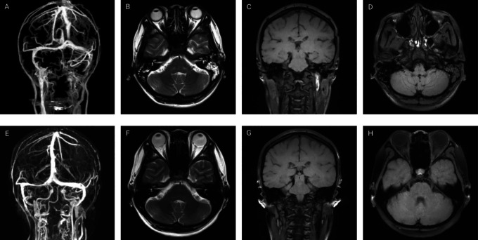
Figure 1.
Neuroimaging of a 33-year-old female with subacute cerebral sinus venous thrombosis. (A) Computed tomography venography demonstrates the absence of flow in the left partial transverse sinus, sigmoid sinuses and internal jugular vein. (B) Axial T2-weighted sequence shows absence of flow-void in the left transverse sinus. (C) Coronal magnetic resonance black-blood thrombus imaging (MRBTI) shows hyperintense signal in the superior segment of left internal jugular vein. (D) Axial MRBTI shows an isointense signal inside the left sigmoid sinuses. Three-dimensional reconstruction magnetic resonance venography (E), axial T2-weighted sequence (F), coronal MRBTI (G) and axial MRBTI (H) at 5 months follow-up demonstrate an almost complete recanalization of the left transverse sinus, sigmoid sinuses and internal jugular vein.

