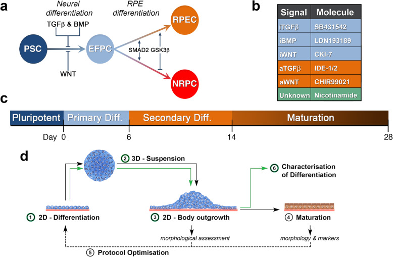
Fig. 1.
A schematic representation for differentiation of PSCs towards retinal cells. a Known signaling pathways and regulators in the differentiation of PSCs to RPE cells (RPECs) and neural retinal progenitor cells (NRPCs) via formation of a common eyefield progenitor cell (EFPC). b List of small molecules for development of a differentiation protocol as substitute endogenous signals: antagonists (blue), agonists (orange). c Timeline for in vitro differentiation of hESC-RPE cells. d Schematic showing stages in hESC differentiation. Stage 1: initiation of differentiation under adherent or 2D conditions. Stage 2: 3D aggregation of differentiating cells forming embryoid bodies. Stage 3: continued differentiation through plating cell bodies for outgrowth of differentiating cells. Stage 4: maturation of RPE cells with identification of cells through morphology and marker expression. Stage 5: optimization of the differentiation protocol through body size, timing of signaling switch and time of plating. Stage 6: characterization of differentiation through gene expression and cell functionality

