Fig. 3.
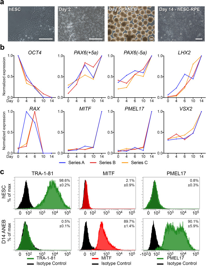
Evidence of in vitro differentiation. a Phase contrast microscopy shows key morphological changes: Human embryonic stem cell (hESC: MEL-1 line) morphology prior to differentiation, formation of differentiating cell monolayers (day 2), and anterior neuroectoderm bodies (ANEBs) (day 5). Plated ANEBs produce expanding sheets of hESC-RPE cell monolayers in the outgrowth by day 14. Scale = 100 μm. b Gene expression over the 14-day differentiation period was measured by qPCR in relation to GAPDH and normalized to maximum expression for each gene over the time period. Normalized expression profiles indicate loss of hESCs (OCT4), upregulation of neuroectoderm [PAX6(+5a), PAX6(−5a), and LHX2], eyefield formation (RAX), and differentiation of RPE cells (MITF and PMEL17). Contaminating neural retinal cell formation was monitored through expression of VSX2. Series A, B, C reflect independent time course experiments and data points reflect the average of technical triplicates. c Flow cytometry indicates expression of TRA-1-81, MITF, and PMEL17 by hESCs at day 0, and differentiation hESC-RPE (ANEBs) at day 14. Data reflect mean + S.D. (n = 3). For each marker, significant differences are detected in the staining of hESCs versus day 14 ANEBs (p < 0.0001)

