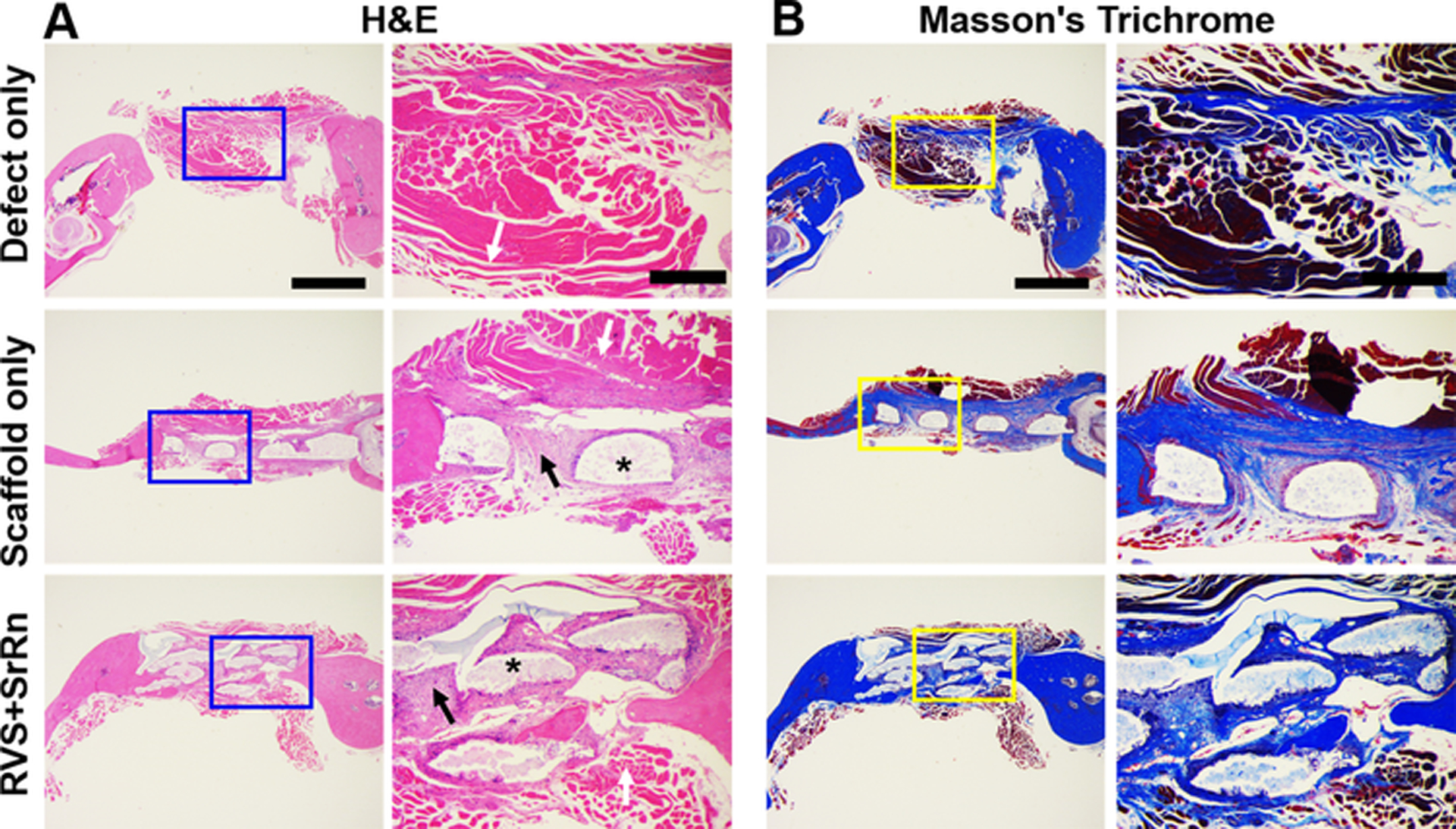
Figure 7.

Histological staining and observations of the newly formed bone in the three groups. (A) H&E staining; (B) Masson’s Trichrome staining. Scale bar: 1 mm for general view, 400 μm for close view. White arrows indicate soft tissue, * indicate scaffolds, black arrows indicate newly formed bones.
