
Figure 4. Increased Scavenger Receptor Expression and Bacterial Uptake in Autophagy-Deficient MEFs.
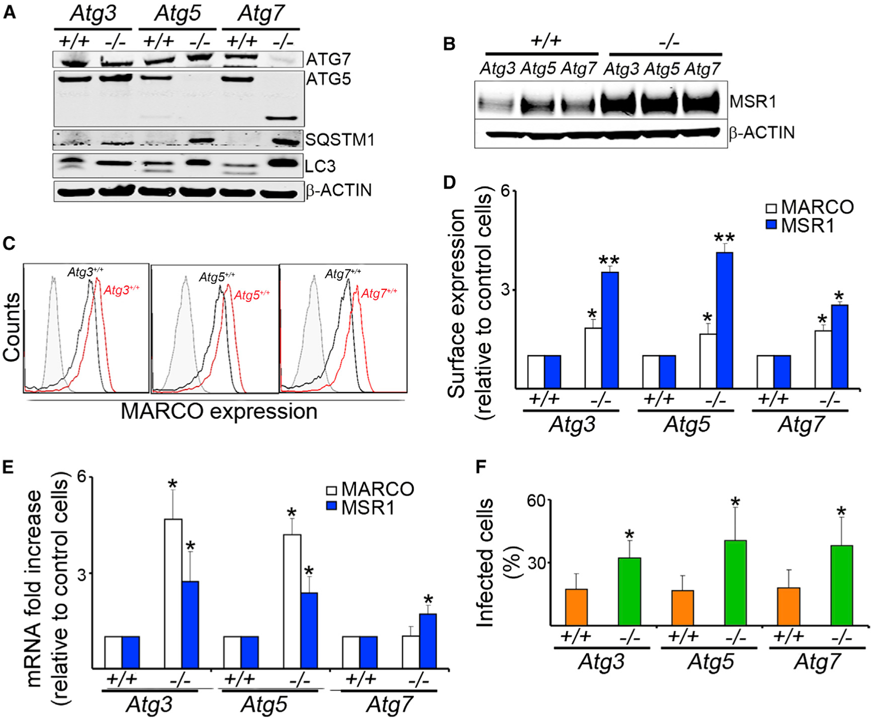
MEFs of Atg3−/−, Atg5−/−, Atg7−/−, and their wild-type controls were evaluated.(A) Antibodies to ATG7, ATG5, SQSTM1, LC3, and b-ACTIN were used to confirm the autophagy-deficient phenotypes.(B) Representative blot of MSR1 and β-ACTIN expression.(C and D) Receptor expression of MARCO and MSR1 was measured by flow cytometry. Representative histograms (C) and quantitative analyses (D) are shown.(E) Real-time PCR was done with mRNA of MEFs and specific primers for MARCO or MSR1.(F) MEFs were infected for 1 hr with GFP-BCG (MOI 10). Percentage of infected cells was determined by immunofluorescence.
Data are mean ± SD, n = 6, *p < 0.05, **p < 0.001.