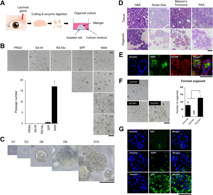
Fig. 1.
Generation of epithelial organoids from human lacrimal glands (LGs). A Scheme for the development and culture of lacrimal tissue-derived organoid. B Optimization of culture media following organoid formation and subculture. Representative images were obtained by light microscopy after culturing for 3 days (Magnification; 10×, Scale bar; 500 µm). C The growth of organoid cultured in optimized LG organoid medium (LGOM). Scale 100 μm. D A comparison of histological analysis between normal lacrimal tissue and formed LG organoid. H&E, Alcian blue, Masson’s trichrome, and PAS staining were performed for identifying the acini structure, mucosubstance secretion, connective tissue, and glycoprotein secretion, respectively (Magnification; 40×, Scale bar; 100 µm). E Confirmation of acinar cells (green; aquaporin 5; AQP5) and epithelial lineage (red; EpCAM) cells in lacrimal tissue (Magnification; 100×, Scale bar; 50 µm). F Organoid formation from EpCAM-positive cell fraction. Representative bright-field images (Magnification; 10×, Scale bar; 500 µm) were obtained after 4 days of cultivation and used for counting the formed organoid (right, graph, N = 4). G Ki67, AQP5, and vimentin (VIM) expression was detected in the organoids and observed using immunofluorescence (Magnification; 100×, Scale bar; 50 µm)

