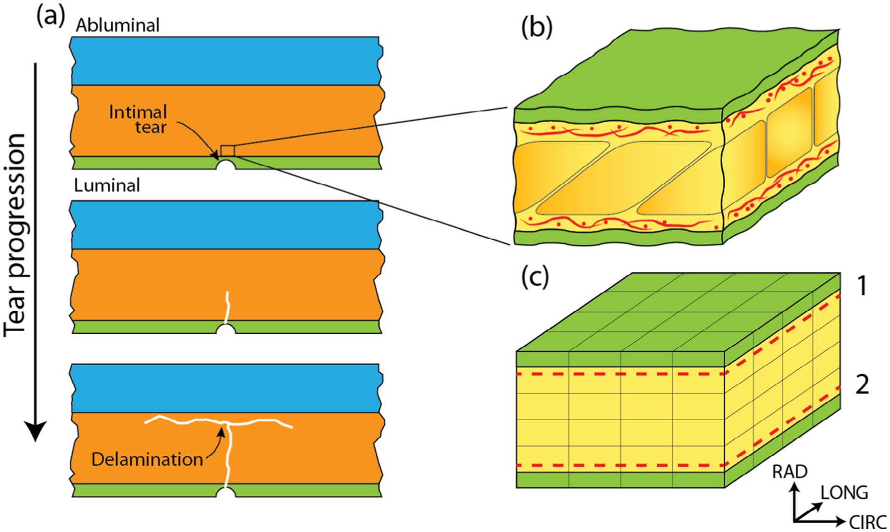
Fig. 1.

(a) The aortic wall is composed of three layers: the intima (green), media (orange), and adventitia (blue). The entire aortic dissection process is confined within the media. Aortic dissection initiates from an intimal tear (top panel), that propagates abluminally in the media (middle panel), and eventually delaminates layers of the media (bottom panel). Media is composed of repeating lamellar units, a representative region of which is shown in (b). The green region denotes the elastic lamellae while the yellow region corresponds to the inter-lamellar space which includes, among other components, collagen fiber networks (red) situated adjacent to the lamellae. A 25 μm × 25 μm × 12.5 μm segment of the representative finite element model of the lamellar unit is shown in (c). As in (b), the green region represents the elastic lamellae and the yellow the inter-lamellar space. The dashed lines show the locations of collagen network layers 1 and 2.
