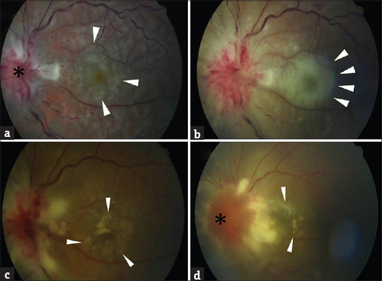
Figure 1.

At the first visit, the fundus revealed significant papillitis (star) with small multifocal grayish yellow lesions (arrow) located mostly in the postequatorial fundus (a). After two days, these grayish lesions became confluent, and a large placoid lesion with typical edge (arrow) was observed mainly within the posterior pole (b). Two weeks after treatment, this confluent grayish placoid lesion was resolved, and multiple yellowish subretinal lesions developed (arrow; c). Four weeks after treatment, these yellowish subretinal lesions diminished (arrow), and papillitis (star) improved (d)
