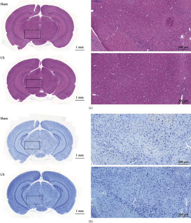
Figure 6.

Histological evaluations of the VTA with and without ultrasound stimulation. (a) Representative H&E staining and (b) Nissl staining of sections from the sham and US groups. The results show no abnormalities (haemorrhaging, tissue damage, or neuron loss) in the VTA.
