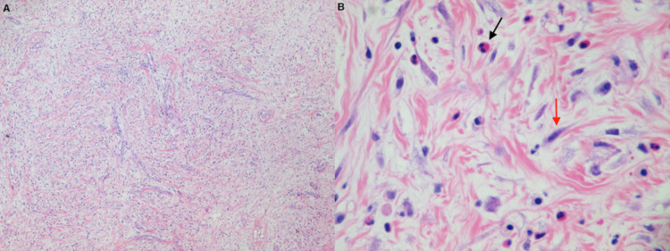
Figure 2. Microscopic appearance of IMT.
A. Low power (×40) H&E staining demonstrating a mild fibroblastic background and collagenous stroma with inflammatory cells consisting of lymphocytes, plasma cells, and scattered eosinophils and neutrophils.
B. High power (x400) microscopic view of H&E stain demonstrating predominance of spindle cells (red arrow) and inflammatory cells (black arrow).
IMT: inflammatory myofibroblastic tumor; H&E: hematoxylin & eosin

