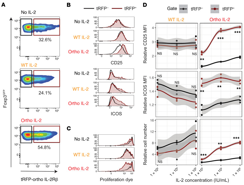
Figure 1. Ortho IL-2 stimulation selectively activates and expands Tregs with oIL-2Rβ.
Tregs transduced with tRFP/oIL-2Rβ were stained with violet cell proliferation dye, followed by incubation with CD3/CD28 activation beads at a cell/bead 1:2 ratio. Flow cytometry analyses performed after 4-day culture are shown. Representative pseudocolor plots (A) and histograms (B and C) of Tregs after incubation with no IL-2 (top), WT IL-2 (middle, 1 × 10³ IU/mL), or ortho IL-2 (bottom, 1 × 10⁵ IU/mL); brown gate: tRFP+ fraction, black gate: tRFP– fraction. (D) Relative ratio to no IL-2 control in CD25 MFI, ICOS MFI, and cell number per well of each gated cell with mean plus 95% confidence intervals at each IL-2 concentration. Quantification of triplicate wells from 1 representative experiment of 3 independent experiments. *P < 0.05; **P < 0.01; ***P < 0.001, calculated between tRFP+ and tRFP–fraction by Welch’s 2-sample t test. WT and ortho IL-2: 1 IU = 312.5 ng.

