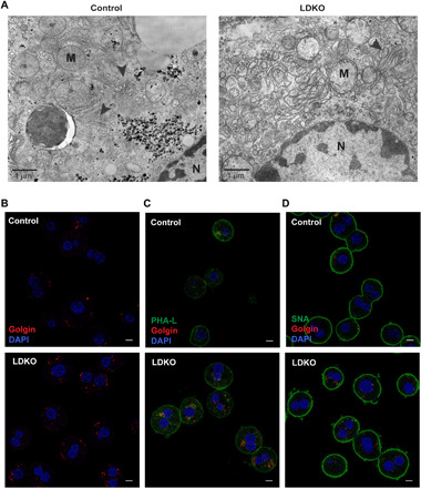
Fig. 5. LDKO hepatocytes exhibit Golgi structural defects.

(A) Electron microscopy imaging shows unstacked Golgi apparatus (arrowheads) in LDKO livers compared to controls. Scale bar, 1 μm. (B) Immunofluorescence staining for a Golgi resident protein, Golgin reveals increased expression in LDKO hepatocytes. DAPI, 4′,6-diamidino-2-phenylindole. This Golgin labeling colocalizes with lectins binding to (C) complex and (D) sialylated glycans in LDKO proteome (hepatocytes cultured from n = 3 mice per group). Scale bars, 10 μm.
