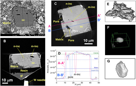
Fig. 1. SEM and XCT slice images of calcite grains, line profiles of RID values, and bird’s-eye views of micrometer-sized inclusions.

(A) A backscattered electron SEM image shows a typical calcite grain (CC-7; see Materials and Methods for the sample number) in matrix. Micrometer-sized inclusions and a large number of nanosized inclusions are observed in an absorption XCT image at 7 keV of CC-7 (B) and a phase-shift image of CC-25 (C). (D) Line profiles of RID values along A-A’ and B-B′ in (C). The RID values larger than zero are due to size effects (see Materials and Methods). Bird’s-eye views of micrometer-sized inclusions obtained from XCT images have a variety of morphologies, from irregular [(E): CC-7], subspherical with facets [(F): CC-27], and tabular hexagonal (negative crystal) [(G): m-inc in (C)]. cc, calcite; m-inc, micrometer-sized inclusion; n-inc, nanosized inclusions; gl, slide glass.
