
Figure 1. Simultaneous calcium imaging and electrophysiology in vivo.
(A) Experimental design. (B) Fluorescence and Vm traces from an exemplar Emx1-s neuron. (C) 5 s of data from the neuron in panel B, showing a 2 AP, a 1 AP, and a 3 AP event. Pre- and post-AP exclusion windows, used to separate events, are illustrated for each event. AP, action potential.
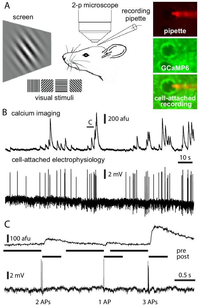
Figure 1—figure supplement 1. Mouse age, cell depth, and firing rate.
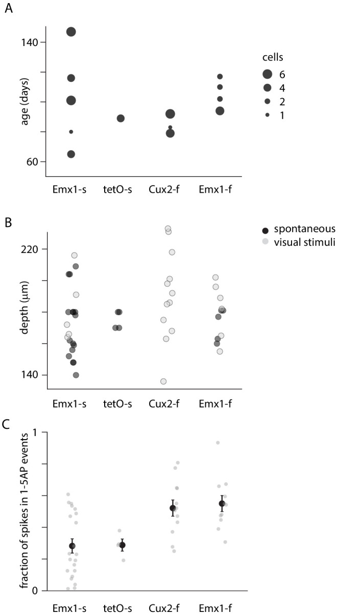
(A) Mouse age. (B) Cell depths. (C) For each neuron, percentage of action potentials (APs) contained within 1–5 AP events.