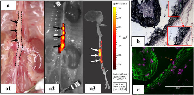
Figure 2.
DiR-NPs targeting in Ang II mouse model. (a) In-vivo targeting study using DiR loaded nanoparticles (DiR-NPs). DiR NPs were injected via tail vein and after 24 h animals were euthanized d to assess the targeting. The signal given by the DiR-NPs in the IVIS images (a2,a3) indicated that DiR-NPs only accumulate at the suprarenal aortic area where the aneurysm developed (a1), suggesting the successful targeting of the nanoparticles to the aneurysmal tissue; (b) VVG staining of the aneurysmal tissue showing elastin damage and (c) fluorescent image of the same site showing distribution of the DiR-NPs (purple) within the tissue, indicating that DiR-NPs target the degraded elastin (green autofluorescence) in the aneurysm.

