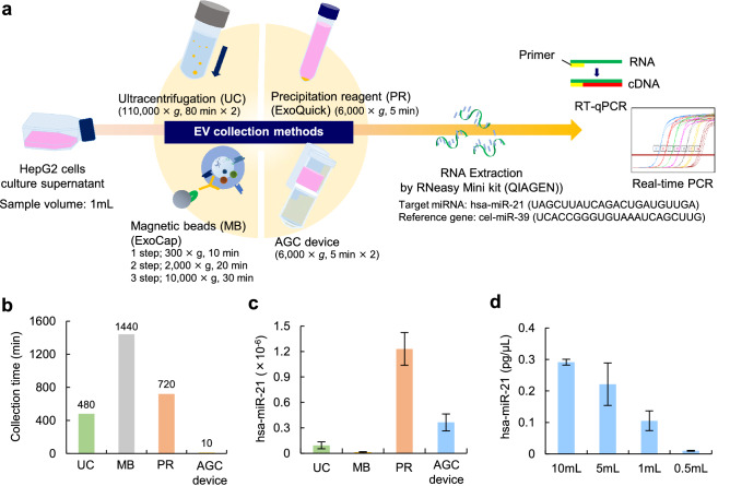
Figure 3.
Comparison of four EV collection methods. (a) A schematic diagram of the real-time PCR analysis of has-miR-21 extracted from HepG2-EVs collected from cell culture supernatant using four EV collection methods (ultracentrifugation [UC], ExoCap [MB], ExoQuick [PR] and the AGC device). (b) The isolation time of HepG2-EVs in UC, MB, PR and the AGC device. (c) The detected level of hsa-miR-21 in UC, PR, MB and the AGC device. (d) The detected level of hsa-miR-21 in the AGC device with different volumes of cell culture supernatant.

