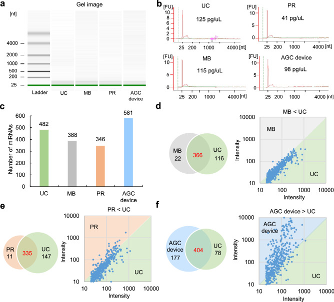
Figure 5.
The miRNA array analysis of EVs collected from human urine. (a) A gel image of nucleotides [nt] of total RNA extracted from EVs collected from human urine using four EV collection methods (UC, MB, PR, and the AGC device) measured by a 2100 Bioanalyzer. (b) The concentration of total RNA extracted from EVs collected from human urine using four EV collection methods (UC, MB, PR, and the AGC device) measured by a 2100 Bioanalyzer. (c) The number of miRNAs extracted from EVs collected from human serum using four EV collection methods (UC, MB, PR, and the AGC device). (d) A venn diagram of the overlap of the number of detected miRNAs between UC (as the standard method) and the other three methods (MB [d], PR [e] and the AGC device [f]).

