
Figure 1.
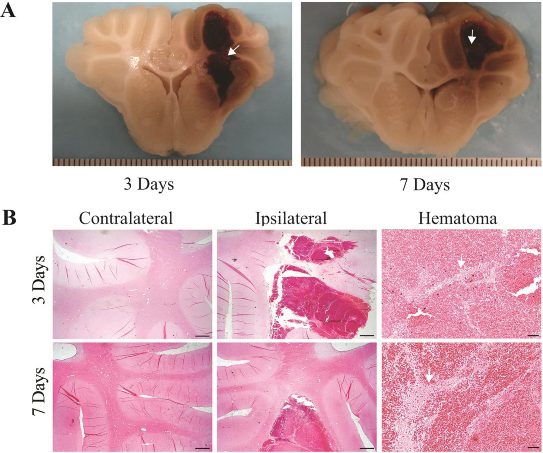
White matter survival in the hematoma. A) Coronal sections of perfused piglet brain showing hematomas with white matter inside (arrows) at 3 and 7 days. Scale intervals in (A) = 1 mm. B) H&E staining of white matter fibers (arrows) inside the hematoma at 3 and 7 days after ICH. Scale bar in the contralateral and the ipsilateral hemisphere = 1 mm; Scale bar in the hematoma = 50 μm.