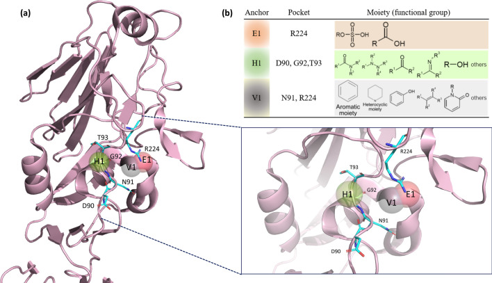
Figure 2.
Receptor-binding site anchors obtained from SiMMap. (a) The RBS structure of the H1N1 HA (chain H of 1RUY) is depicted as a cartoon and three anchors as transparent spheres. Red E1 stands for the electrostatic force, green H1 for hydrogen bond force, and grey V1 for van der Waals forces. The corresponding binding pockets (residues) are shown in the cyan sticks. (b) The table presents binding pockets and moieties for each anchor. Each moiety of the anchor represents the functional group preference of the top-ranked compounds.

