Figure 1.
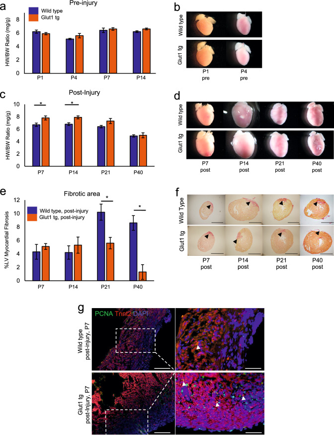
Increase in intracellular glucose promotes cardiac regeneration in Glut1 transgenic heart. (a) Heart weight-body weight (HW/BW) ratio of Wild type and Glut1 transgenic (Glut1 tg) mice without injury. n = 3–7 for each group, p = n.s. (b) Representative images of hearts. (c) Heart weight-body weight (HW/BW) ratio of Wild type and Glut1 transgenic mouse post-injury. The hearts were cryoinjured at P1 and examined at P7, 14, 21 and 40. Note that the HW/BW is higher in Glut1 transgenic hearts at P7 and 14. n = 4–9 for each group, *p < 0.05. (d) Images of representative hearts post-injury. Note the ballooning of the heart at P14 in both wild type and Glut1 transgenic hearts. (e) %Fibrotic area measured by Image J capture of Picrosirius red stainings of the hearts. Hearts were cryoinjured at P1 and examined at P7, 14, 21 and 40. n = 5–8 for each group, *p < 0.05. (f) Representative images of Picrosirius red staining of wild type and Glut1 transgenic hearts. Arrowheads indicate fibrotic area. Scale bar = 200 µm. (g) PCNA staining of the sections from wild type and Glut1 transgenic hearts 7 days post-injury. Sections were stained with a cardiac marker (Tnnt2; Red), proliferation marker (PCNA; Green) and a nuclear marker (DAPI; Blue). Note that PCNA staining is more abundant in Glut1 transgenic heart. Arrowheads indicate PCNA positive cardiomyocytes. Scale bar = 50 µm and 20 µm respectively.

