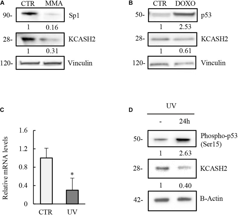
FIGURE 5.
Modulation of KCASH2 expression by p53 and Sp1 is conserved in mouse. (A) Inhibition of Sp1 leads to a decrease of KCASH2 protein levels. MEF cells were treated with MMA (500 nM) for 24 h. Protein lysates were analyzed by Western Blot, using Sp1 and KCASH2 antibodies. Anti-vinculin was used as loading control. (B–D) p53 downregulates KCASH2 expression. (B) MEF cells were cultured with DOXO (5 μM) for 10 h, then cells lysates were analyzed by Western blot. Vinculin was used as loading control. (C,D) MEF cells were exposed to UV-C light and collected after 24 h. Then, RT-qPCR analysis was performed (C). Endogenous KCASH2 mRNA levels were normalized on GAPDH and HPRT, and represented as fold-induction on CTR. Data are representative of three experiments and presented as mean ± SD. ∗p < 0.05. Endogenous KCASH2 protein and p53 activation (D) were analyzed by Western blot and revealed with antibodies against KCASH2 and Ser15 phosphorylated p53. Anti-β-actin is shown as loading control.

