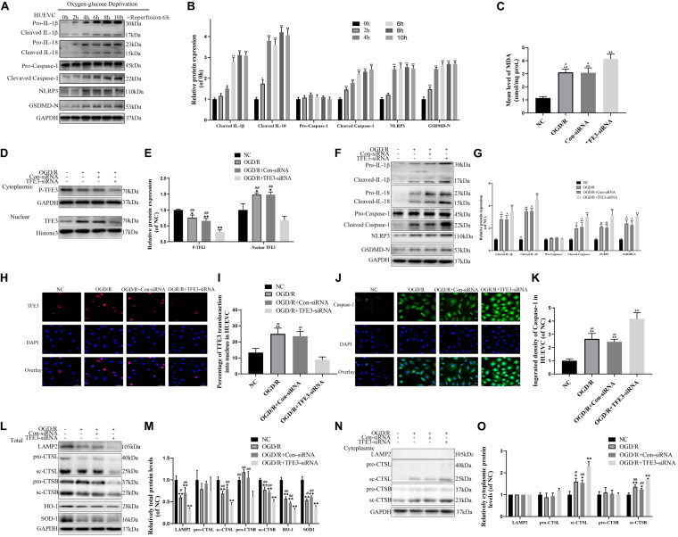
FIGURE 6.
TFE3 silencing aggravates lysosomal malfunction and promotes pyroptosis in the OGD/R cell model. (A) Western blotting of the pyroptosis markers in HUVECs. (B) Quantification of pyroptosis related proteins immunoblots in HUVECs. (C) ELISA of the oxidation product MDA in HUVECs from NC, OGD/R, OGD/R + Con-siRNA, and OGD/R + TFE3-siRNA groups. (D) Western blotting of the level of cytoplasmic and nuclear TFE3 in HUVECs. (E) Quantification of TFE3 immunoblots. (F) The protein levels of pyroptosis related proteins in HUVECs from each group were detected via western blotting. (G) Quantification of pyroptosis related proteins. (H) Immunofluorescence was performed to exhibit the nuclear translocation of TFE3 (red) in the HUVECs (scale bar: 25 mm). (I) Percentage of TFE3-positive cells. (J) Immunofluorescence to detect the caspase-1 expression level was conducted to exhibit the pyroptosis level in the HUVECs (scale bar: 25 mm). (K) The fluorescence intensity of caspase-1 analyzed using ImageJ. (L,N) Western blotting of LAMP2, CTSL, CTSB, SOD1, and HO-1 in HUVECs from each group. (M,O) Western blot and its quantification showed the level of LAMP2, CTSL, CTSB, SOD1, and HO-1. Data are the mean ± SEM, n = 6 per group. *p < 0.05 and **p < 0.01, vs. NC group. #p < 0.05 and ##p < 0.01, vs. OGD/R + TFE3-siRNA group.

