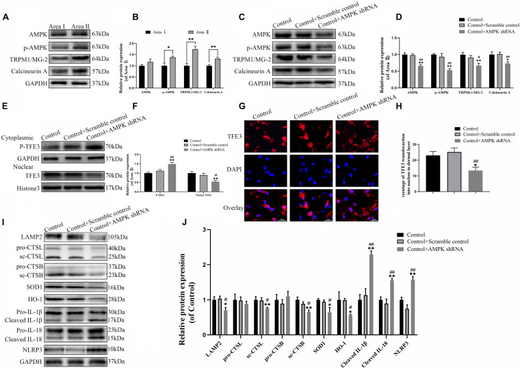
FIGURE 7.
The activity of TFE3 is regulated by AMPK-MCOLN1-calcineurin signaling pathways. (A,C) Western blotting of AMPK, p-AMPK, TRPML1/MG-2, and calcineurin in flap tissue. (B,D) Quantification of AMPK, p-AMPK, TRPML1/MG-2, and calcineurin immunoblots. (E) Western blotting of cytoplasmic TFE3 and nuclear TFE3 in flaps. (F) Quantification of cytoplasmic TFE3 and nuclear TFE3 immunoblots. (G) Immunofluorescence of nuclear translocation of TFE3 in flaps (scale bar: 25 mm). (H) Percentage of TFE3-positive cells. (I) Western blotting of LAMP2, CTSL, CTSB, SOD1, HO-1, cleaved-IL-1β, cleaved-IL-18, and NLRP3. (J) Histograms of the optical density values of LAMP2, CTSL, CTSB, HO-1, SOD1, cleaved-IL-1β, cleaved-IL-18, and NLRP3. Data are the mean ± SEM, n = 6 per group. *p < 0.05 and **p < 0.01, vs. control or area II group. #p < 0.05 and ##p < 0.01, vs. control + scramble control group.

