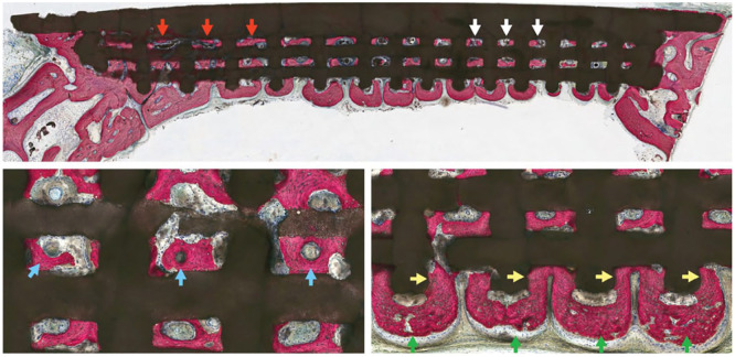
Figure 3.
Dipyridamole coated β-TCP scaffold assessment: quadrant scaffold demonstrates bone regeneration through scaffold porosity, at both larger (red arrows) and smaller (white arrows) pore dimensions. (Below, left) Highly cellular and vascularized bone formation is seen within scaffold interstices. Intramembranous-like healing is observed with regions of mature, lamellar-like bone formation (blue arrows). (Below, right) Bone formation is guided by highly osteoconductive scaffold dimensions as new bone formation is directed from scaffold pore-to-pore (green arrows) while interacting with scaffold struts (yellow arrows). Adapted from Maliha et al.18

