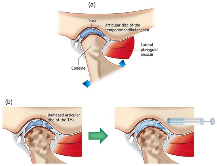
Figure 5.
(a) Schematic image of the anatomical structure of temporomandibular joint (TMJ) and the most common target sites for treating temporomandibular disorder (TMD). The image shows components of normal joint anatomy, including the articular disk of TMJ, mandibular fossa, the head of the mandibular condyle, lateral pterygoid muscle, and TMJ capsule enclosing the disk. (b) TMD morphology; the head of the mandibular condyle and the articular disk lose their structures and functions. Intra-articular injection: injection with syringe and needle can deliver proper biomolecules into TMJ capsule for treating TMD as adapted from Dashnyam et al.81

